Atlas of Corneal Imaging 1st Edition – PDF/EPUB Version Downloadable
Author(s): J. Bradley Randleman
Publisher: CRC Press
ISBN: 9781041256458
Edition: 1st Edition
$49,99
Delivery: This can be downloaded Immediately after purchasing.
Version: Only PDF Version.
Compatible Devices: Can be read on any device (Kindle, NOOK, Android/IOS devices, Windows, MAC)
Quality: High Quality. No missing contents. Printable
Recommended Software: Check here

Important: No Access Code
Description
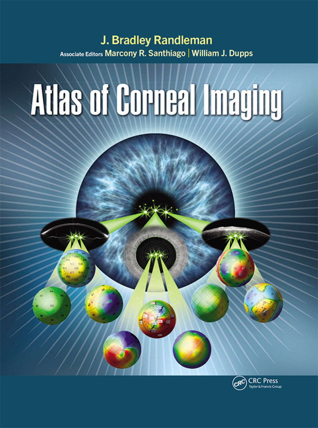
A comprehensive reference for physicians, surgeons, and trainees, Atlas of Corneal Imaging covers all aspects of corneal imaging from basic map interpretation to advanced diagnostic uses and features over 1200 illustrative images and figures representing a wide variety of devices and techniques. Drs. J. Bradley Randleman, Marcony Santhiago, and William J. Dupps Jr guide readers through the process of analyzing corneal images using a multitude of different techniques, technologies, and individual devices. This creates a complete picture of the cornea’s basic methods and pathological processes and allows readers to directly visualize how their technology would display the pathology in question. Atlas of Corneal Imaging is designed to help practitioners recognize subtle findings and evaluate signs of weakening or pathology, no matter how they present or what device is being used. Multiple iterations of the same clinical condition are shown with numerous complementary images for the same eye to provide a comprehensive presentation of each case. Chapters feature information on: Topographic patterns and mapping Corneal ectasia evaluations Cornea and refractive surgery evaluations Clinical correlations with corneal disorders Cornea and refractive surgery complications Evaluation for cataract surgery Atlas of Corneal Imaging fills a significant void in corneal imaging resources available today by presenting an image-first approach to understanding all the many different technologies for imaging the cornea.