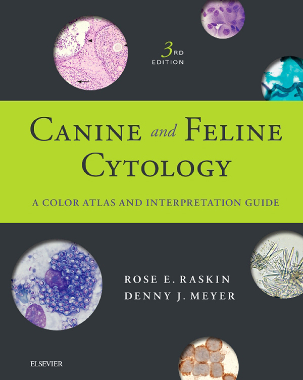
Canine and Feline Cytology A Color Atlas and Interpretation Guide 3rd Edition – PDF/EPUB Version Downloadable

Author(s): Rose E. Raskin; Denny Meyer
Publisher: Saunders
ISBN: 9781455740833
Edition: 3rd Edition

$49,99

Delivery: This can be downloaded Immediately after purchasing.
Version: Only PDF Version.
Compatible Devices: Can be read on any device (Kindle, NOOK, Android/IOS devices, Windows, MAC)
Quality: High Quality. No missing contents. Printable

Recommended Software: Check here

Important: No Access Code

Description

“This is a ‘go-to’ reference text for a serious cytologist. ” Reviewed by: Kathleen Tennant on behalf of Veterinary Record, November 2015

  • Comprehensive coverage of all body systems and body fluids — and the pathological changes associated with various infectious agents — emphasizes areas in which the application of cytology has the greatest diagnostic value.
  • Exceptional-quality, full-color photomicrographs show both normal and abnormal tissue and also include detailed legends.
  • Discussions of clinical, differential, and cytological diagnosis accompany the illustrations of lesions and conditions in each chapter.
  • Helpful hints for improving specimen quality are provided in discussions of common errors and problems encountered in the preparation of cytological specimens.
  • Coverage of histology in organ system chapters demonstrates the histological or histopathologic corollary of cytologic findings.
  • Clear, concise descriptions include sampling techniques, slide preparation and examination, and guidelines for interpretation, leading to accurate in-house and commercial laboratory diagnosis.
  • Easy-to-use, well-organized format includes many tables, algorithms, boxes, and Key Point callouts for at-a-glance reference.

  • NEW examples and identifiers and a new discussion on artifacts address the latest developments in canine and feline cytology.
  • NEW! Dozens of new images include photomicrographs of blood and bone marrow cytology.
  • NEW! All-new appendix summarizes clinically relevant information on artifacts, special tests, specific characteristics, and much more.
  • UPDATED sampling techniques and examination guidelines include information on how and where to test for specific conditions.